College of Optical Sciences

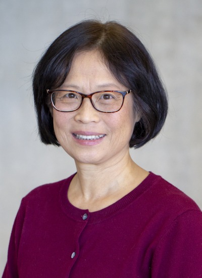
Linan Jiang

Associate Research Professor, Aerospace and Mechanical Engineering
Associate Research Professor, Optical Sciences
Contact
(520) 621-6120

Work Summary

Dr. Jiang is an Associate Research Professor with the Department of Aerospace and Mechanical Engineering, College of Engineering, University of Arizona. Dr. Jiang develops integrated microsystems, including microfluidics, Lab-on-Chip, Organs-on-Chips, microOptics, flexible optical interconnect and microsensors.

Research Interest

Dr. Jiang’s research interests broadly lies in micro/nano technology applications to advance engineering, manufacturing, biology, and medicine. One of Dr. Jiang's research focus is development of human models of Organ(s)-on-Chips in collaboration with multidisciplinary teams of colleagues. These Organs-on-Chip microsystems closely mimic in vivo environments, implement an integrated approach and enable investigations in a well-controlled manner. They are used to study roles of tissue/organ interactions, mechanisms in tumor progression, invasion, metastasis, and dormancy, as well as disease treatments for targeted therapy and personalized medicine.

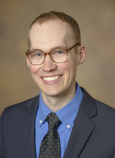
Travis William Sawyer

Assistant Professor, Biomedical Engineering
Assistant Professor, Optical Sciences
Research Assistant Professor, Medical Imaging
Member of the Graduate Faculty
Primary Department
Contact
520-621-8068

Work Summary

Travis Sawyer is an Assistant Professor of Optical Sciences and Health Sciences. His laboratory focuses on research to develop new imaging techniques and technologies for the early detection of cancers including ovarian, skin, esophageal, and stomach cancers. He also has extensive experience in image processing, having started an independent company prior to joining the faculty in Optical Sciences. Travis supports science outreach to encourage younger generations to pursue careers in STEM.

Research Interest

Travis Sawyer is an Assistant Professor of Optical Sciences and Health Sciences. He received his BS in Optical Sciences from the UA (2017) before attending the University of Cambridge to receive his MPhil in Physics (2018). He then returned to the UA pursue his PhD in Optical Sciences (2021) where he focused on developing novel imaging techniques for ovarian cancer detection. After graduating, he joined the faculty at the College of Optical Sciences to establish the Biomedical Optics and Optical Measurement Lab. His research interests include gastrointestinal cancer detection, where he develops endoscopes incorporating optical coherence tomography, fluorescence imaging, and other novel imaging modalities, with a focus on image analysis through machine learning techniques. He has extensive experience in animal and human studies, as well as optical design and has collaborated with the University of Cambridge to develop endoscopes using hyperspectral and phase imaging. Previously, he developed visual recognition software for detailed image capture, enabling discoveries in astronomy, art preservation, and the biomedical sciences. Travis is also a strong supporter of science outreach, regularly organizing and supporting efforts to educate the community and encourage careers in Science.

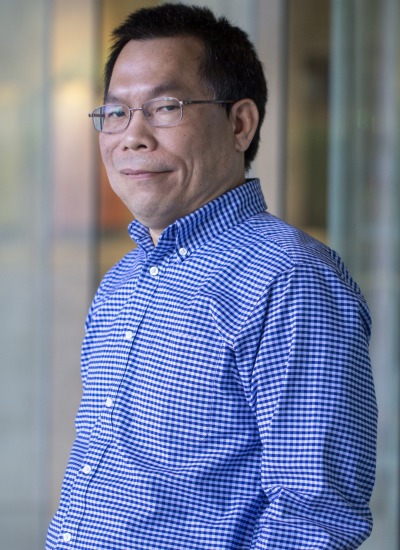
Rongguang Liang

Professor
Professor, BIO5 Institute
Member of the Graduate Faculty
Primary Department
Contact
520-621-4995

Work Summary

Dr. Liang’s research focuses on three major fields: biomedical imaging, advanced imaging, and optical engineering. In biomedical imaging, his lab has been working on various imaging techniques for image guided surgery, cancer detection and diagnosis, including low-cost, mobile imaging platform for oral cancer screening in low-resource setting and multimodal OCT system for precision oral cancer detection and diagnosis. His lab has also been investigating new optical techniques for tissue imaging, such as confocal, light sheet, multiphoton, and hyperspectral imaging. In advanced imaging, Dr. Liang’s lab aims to extract additional information based on phase, polarization, and spectrum. His lab has developed snapshot imaging techniques for polarization and spectral imaging. In optical engineering, Dr. Liang’s lab is known for the capabilities in optical design, fabrication, and testing. His group has been developing compact optical systems for medical, consumer, industrial, and space applications, including three cameras used in NASA’s OSIRIX-REx mission. In addition to precision diamond turning, his lab has developed unique 3D printing method for fabricating precision freeform optics. Dr. Liang’s group has also been very active on optical metrology, with a focus on freeform and snapshot metrology.

Research Interest

Dr. Liang’s research interest is to develop, prototype, and evaluate advanced optical imaging techniques for medical, consumer, and industrial applications.


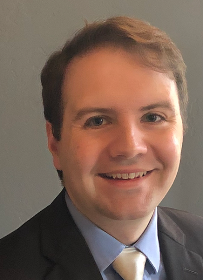
Euan Mcleod

Associate Professor, Optical Sciences
Member of the Graduate Faculty
Assistant Professor, BIO5 Institute
Primary Department
Contact
(520) 621-6997

Work Summary

Prof. Euan McLeod studies optical systems for sensing and imaging objects at the nanoscale. His lab uses optical tweezers to "print" photonic devices at higher resolution out of more types of materials than other 3D printers. Euan also works on cost-effective field-portable lensfree holographic microscopes that provide high resolution across an ultra-large field of view. These microscopes are used for biomedical sensing and environmental air quality monitoring.

Research Interest

Euan McLeod, Ph.D., works at the intersection of nanophotonics, soft materials science, and many-body systems. One of his current major research thrusts is to use optical tweezers combined with biomolecular functionalization to assemble nanostructured 3D devices out of colloidal nanoparticle building blocks. Euan also works on developing lensfree holographic microscopes that provide high resolution across an ultra-large field of view in cost-effective and compact platforms. Euan is developing new methods to improve the resolution and sensitivity of these microscopes to sense ultrafine nanoparticles like aerosols and viruses. By combining these microscopes with microfluidic chambers, he is working to develop highly multiplexed biomedical sensors. All of these areas of experimental research are supported by extensive computational and theoretical efforts. Previously in his career, Euan has published extensive research in high-speed acoustic lensing, laser-materials processing at the nanoscale, and free-surface microfluidic instabilities.

Judith Su

Assistant Professor, Biomedical Engineering
Assistant Professor, Optical Sciences
Assistant Research Scientist, Chemistry and Biochemistry
Assistant Professor, BIO5 Institute
Primary Department
Contact
(520) 621-4240

Research Interest

Judith Su is an Assistant Professor in Biomedical Engineering and an Assistant Professor of Optical Sciences at the University of Arizona. She is also an Associate Member of the University of Arizona Cancer Center. Judith received her B.S. and M.S. from MIT in Mechanical Engineering and her Ph.D. from Caltech in Biochemistry & Molecular Biophysics. Her background is in imaging, microfabrication, and optical instrument building for biological and medical applications. In general, her research interests are to develop new imaging, sensing, and rheological techniques to reveal basic biological functions at the molecular, cellular, and tissue levels. Recently her work has centered on label-free single molecule detection using microtoroid optical resonators with a focus on basic research, and translational medicine through the development of miniature field portable devices.

Matthew A Kupinski

Professor, Optical Sciences
Professor, Radiology
Professor, Applied Mathematics - GIDP
Professor, BIO5 Institute
Primary Department
Department Affiliations
Contact
(520) 621-2967

Work Summary

Matthew Kupinski works in diverse areas of imaging including x-ray, gamma-ray, diffuse optical, magnetic resonance, and neutron imaging.

Research Interest

My research interests are in the field of theoretical image science with emphasis on medical imaging. I currently study task-based measures of image quality in which one must define the task the images are to be used for and the observer who will be performing this task, to properly measure and optimize the quality of images and imaging systems. We take the stance that imaging systems should be designed to best enable the observer to detect tumors and not base design decisions on resolution, contrast, etc. Topics in my research group include accurate system modeling, statistical modeling, observer performance metrics, signal-detection theory and general image science.

Dongkyun Kang

Assistant Professor, Optical Sciences
Assistant Professor, Biomedical Engineering
Assistant Professor, BIO5 Institute
Primary Department
Contact
(520) 621-6997

Work Summary

We are developing low-cost in vivo microscopy devices that can visualize cellular details of human tissues in vivo and help disease diagnosis and treatment in low-resource settings, high-speed tissue microscopy technologies that can examine entire organ under risk of having malignant diseases and detect small, early-stage lesions, and miniature microscopy devices that have the potential to examine anatomically-challenging human organs and facilitate integration of microscopic imaging with other imaging modalities.

Research Interest

My research is focused on developing novel optical microscopy technologies and improving patient care using these technologies. My research area includes (1) low-cost smartphone in vivo microscopy, (2) high-speed comprehensive in vivo endomicroscopy, and (3) ultraminiature endomicroscopy. (1) Low-cost smartphone in vivo microscopy: I am currently leading a NIH-sponsored research project for developing smartphone confocal microscope and diagnosing Kaposi's sarcoma in Uganda with the smartphone confocal microscope. I will further advance the smartphone microscopy technology and address other applications, including diagnosis of cervical and oral cancers in low-resource settings, large-population screening of skin cancers in the US, and aiding science and medical educations. (2) High-speed comprehensive in vivo endomicroscopy: I have previously developed a high-speed confocal microscopy system and endoscopic imaging catheters and acquired largest in vivo confocal images of human organ reported. At the UA, I plan to further advance the technology by i) increasing the imaging speed by orders of magnitude and ii) incorporating fluorescence imaging modality. (3) Ultraminiature endomicroscopy: In my previous research, I have developed miniature endoscopic catheters that can visualize internal organs in vivo through a needle-sized device. At the UA, I will develop microscopic imaging catheter with a extremely small diameter and utilize it for guiding cancer diagnosis and treatment.