Anatomy

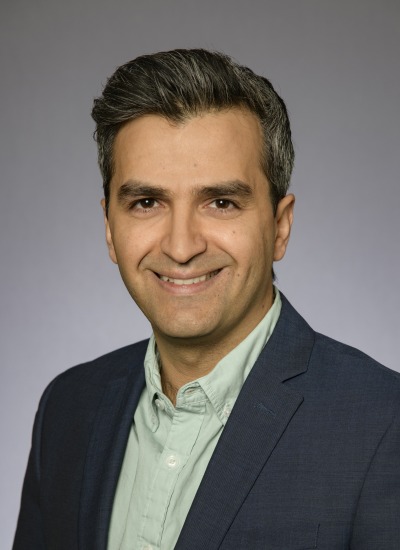
Qin Chen

Director, Pharmacogenomics
Professor, Pharmacogenomics
Professor, Pharmacology and Toxicology
Professor, Pharmacology
Professor, Applied BioSciences - GIDP
Associate, Center for Toxicology
Member of the Graduate Faculty
Professor, Cancer Biology - GIDP
Professor, Genetics - GIDP
Endowed Professor, Holslaw - Pharmacogenomics
Professor, BIO5 Institute
Primary Department
Contact
(520) 626-9126

Research Interest

Our laboratory studies cellular and tissue injury due to oxidative stress. We pioneered the discovery that cells surviving oxidative stress develop hypertrophy. This discovery has been validated in many cell types as a consequence of cellular stress and survival response. Enlarged cells contribute to loss of functionality during the development of diseases. In the myocardium, cardiomyocyte hypertrophy can be detected as a result of ischemic injury and contributes to heart failure. Continuing on the investigation of mechanisms of cell survival has led us to focus on cellular defense system. From our many years of comprehensive and systematic studies on cellular and molecular events initiated by oxidative stress, Nrf2 stands out as the key controller for cell defenses. We have made several discoveries in recent years, including 1) oxidative stress induced de novo Nrf2 protein translation; 2) Nrf2 physically interacts with mitochondria and protects mitochondria against oxidative stress induced decay; and 3) deficiency in Nrf2 sensitizes the myocardium to ischemic injury.

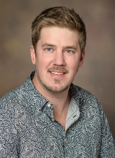
Kaveh Laksari

Assistant Professor, Biomedical Engineering
Assistant Professor, Aerospace-Mechanical Engineering
Member of the Graduate Faculty
Assistant Professor, BIO5 Institute
Primary Department
Department Affiliations

Work Summary

https://www.engr.arizona.edu/~klaksari/

Research Interest

Our research focuses applying engineering methods to problems in biology with the goal of improving human health. In particular we aim to understand the underlying mechanisms of traumatic brain injury in order to better prevent and diagnose. We also research on the cerebral hemodynamics and the effect it can have on neurodegenerative diseases and stroke. We use an array of computational and experimental approaches including finite element modeling, magnetic resonance imaging and impact biomechanics. https://www.engr.arizona.edu/~klaksari/

Brett Colson

Assistant Professor, Cellular and Molecular Medicine
Assistant Professor, Physiological Sciences - GIDP
Assistant Professor, Biomedical Engineering
Assistant Professor, Clinical Translational Sciences
Member of the Graduate Faculty
Assistant Professor, BIO5 Institute
Primary Department
Contact
(520) 621-1950

Research Interest

The research goal of my laboratory is to understand the molecular motions and interactions of proteins involved in regulating contractile function of healthy cardiac and skeletal muscle, to determine the culprits of contractile dysfunction and remodeling in muscle disorders and cardiovascular disease, and then apply these insights to design novel therapies. We use biophysical approaches, such as time-resolved spectroscopy with site-directed probes to assess protein structural dynamics and mechanical measurements of isolated muscle fibers to assess contractile force and kinetics, in order to establish structure-function relationships inherent to the molecular, biochemical, and physiological mechanisms.

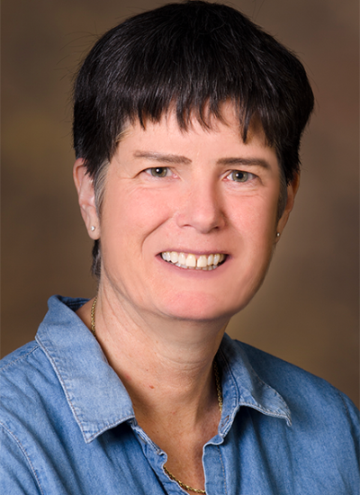
Daniela C Zarnescu

Professor, Cellular and Molecular Medicine
Professor, Neuroscience
Professor, Applied BioSciences - GIDP
Professor, Neurology
Professor, Genetics - GIDP
Professor, Molecular and Cellular Biology
Professor, Neuroscience - GIDP
Member of the Graduate Faculty
Professor, BIO5 Institute
Primary Department
Contact
(520) 626-1478

Work Summary

We are working to uncover the molecular mechanisms of aging and neurodegenerative diseases using a combination of genetic, computational and pharmacological tools, and a diverse array of experimental models. We also seek to develop therapies for ALS and related neurodegenerative diseases.

Research Interest

Fragile-X syndrome, which includes mental and physical defects and is the most common form of inherited mental retardation. Keywords: Neurodegeneration, ALS, Aging

Stephen H Wright

Professor, Physiology
Professor, Biochemistry/Molecular Biophysics
Professor, Physiological Sciences - GIDP
Investigator, Center for Toxicology
Professor, BIO5 Institute
Primary Department
Department Affiliations
Contact
(520) 626-4253

Work Summary

The kidney plays a critical role in clearing the body of potentially harmful compounds, including many commonly prescribed drugs. Unfortunately, this also sets the kidney up as a site where multiple drugs can interact in unwanted ways. We study the cellular transport processes responsible for renal drug clearance with the intent of developing predictive models that can assist clinicians, drug companies, and the Food & Drug Administration in their efforts to increase patient safety.

Research Interest

Stephen Wright, PhD, is focused on understanding the molecular and cellular physiology of organic electrolyte transport in the kidney. The kidney, particularly the proximal tubule, actively secretes a wide array of organic ions, largely derived from dietary or pharmaceutical sources. Many of these compounds are toxic and renal secretion of these xenobiotic compounds plays a critical role in protecting the body from these agents. However, this task also places the kidney in harm's way, and the development of nephrotoxicity is one consequence of the renal secretion of what are typically referred to as organic anions and organic cations. Dr. Wright’s lab currently studies the renal transport of organic anions and cations at several different levels of biological organization.At the molecular level, they clone individual transport proteins for use in studies that gauge the effect of protein and substrate structure on the transport process. At the cellular level, Dr. Wright and his lab use cultured cells (including primary renal cells, continuous renal cell lines, and generic cells lines for the expression of cloned transport proteins) in studies of the activity and regulation of transport activity. At the tissue level, they use isolated, intact renal proximal tubules, including single non-perfused and perfused tubules, to study the process of organic electrolyte secretion as it occurs in the native renal epithelium.Studies employ a wide array of methodologies, including molecular cloning, site-directed mutagenesis, construction of fusion proteins, kinetic assessment of membrane transport in cultured cells, suspensions of isolated renal tubules and in single tubule segments using radiometric and real-time optical approaches, computationally-based assessment of transporter, and substrate structure and 3D distribution of cell type distribution along the renal nephron. Keywords: Membrane Transport; Kidney; Drug Clearance

Jean M Wilson

Professor, Cellular and Molecular Medicine
Director, Willed Body Program
Professor, Cancer Biology - GIDP
Professor, Neuroscience - GIDP
Professor, BIO5 Institute
Member of the General Faculty
Member of the Graduate Faculty
Primary Department
Contact
(520) 626-2557

Research Interest

Jean M. Wilson, Ph.D. is a Professor of Cellular and Molecular Medicine at the University of Arizona and member of the Arizona Cancer Center. Dr. Wilson’s work focuses on the establishment and maintenance of the mucosal barrier of the intestine. The cells of the intestine provide a selective barrier to pathogens and toxins, and loss of this barrier function is fundamental to pathologies such as inflammatory bowel disease and bacterial infection. In addition, loss of cellular interactions important for barrier function may predispose these cells to cancer. Work in Dr. Wilson’s laboratory focuses on a protein that is highly expressed in developing intestine, implying a critical role in the formation of the intestinal epithelium. Disruption of this protein compromises junctional integrity and epithelial polarity. Furthermore, expression of this protein is decreased in a model of neonatal necrotizing enterocolitis, a disease of newborns with high morbidity and mortality. These findings implicate this protein in the maintenance of intestinal barrier function in the neonate. In addition, continued expression in the adult intestine positions it to regulate epithelial permeability and polarity throughout life. Our studies focus on protein partners that interact with this protein with the goal of identifying the molecular machinery that regulates this pathway.

Jennifer A Teske

Associate Professor
Associate Professor, Neuroscience - GIDP
Associate Professor, Physiological Sciences - GIDP
Member of the General Faculty
Member of the Graduate Faculty
Primary Department
Department Affiliations
Contact
(520) 621-3081

Research Interest

Dr. Jennifer Teske, PhD is an Assistant Professor in the Department of Nutritional Sciences. Her primary research interest is the study of the metabolic consequences of environmental noise stress as it relates to the whole-organism stress response and human health.

Timothy W Secomb

Professor, Physiology
Professor, Biomedical Engineering
Professor, Mathematics
Professor, Applied Mathematics - GIDP
Professor, Physiological Sciences - GIDP
Research Professor, Arizona Research Labs
Professor, BIO5 Institute
Contact
(520) 626-4513

Research Interest

Timothy Secomb, PhD, studies the microcirculation, a network of extremely small blood vessels that supply oxygen and nutrients to all parts of our tissues. The focus of work in his research group is the use of mathematical and computational approaches to study blood flow and mass transport in the microcirculation. Working in collaboration with experimentalists, the aim is to understand quantitatively the processes involved. Dr. Secomb examines the relationship between red blood cell mechanics and flow resistance in microvessels. Theoretical predictions agree well with observations in glass tubes, but resistance is higher living tissue. The major cause is the presence of a relatively thick macromolecular lining (endothelial surface layer) on the walls of microvessels. He also simulates oxygen exchange between networks of microvessels and surrounding tissues in skeletal muscle and tumors. In skeletal muscle, oxygen can be exchanged diffusively between arterioles and capillaries, and Dr. Secomb’s lab is studying the determinants of maximal oxygen consumption. In tumors, the relationship between network structure and occurrence of local hypoxic (radiation-resistant) regions is a source of curiosity. They are analyzing the delivery of chemotherapeutic drugs in tumor tissues, and developing improved models to describe the responses of tumor cells to chemotherapy and radiation. Models for the structural responses of microvessels to functional demands are being developed. Maintenance of a stable, functionally adequate distribution of vessel diameters can be achieved if each vessel responds to changes in wall shear stress, intravascular pressure and local metabolic conditions, and if mechanisms exist for information transfer upstream and downstream along flow pathways. Models for the active regulation of blood flow by changes in vascular tone are also being developed, taking into account vascular responses to wall shear stress, pressure and local metabolic state, and including effects of conducted responses along vessel walls. Another project in the group is the development of computer simulations for the dynamics of the left ventricle that can be run in real time and provide a tool for analysis of data derived from ultrasound echocardiography images.

Gregory C Rogers

Professor, Cellular and Molecular Medicine
Associate Professor, Cellular and Molecular Medicine
Associate Head, Faculty Development
Associate Professor, Cancer Biology - GIDP
Associate Professor, Molecular and Cellular Biology
Associate Professor, Genetics - GIDP
Associate Professor, BIO5 Institute
Primary Department
Contact
(520) 626-3925

Research Interest

Gregory C. Rogers, PhD, is an Assistant Professor at the University of Arizona College of Medicine in the Department of Cellular and Molecular Medicine. His laboratory is located in the University of Arizona Cancer Center.The Rogers laboratory is interested in the molecular mechanisms cells use to maintain stability of their genomes. This is medically relevant because genomic instability can promote tumorigenesis. During mitosis, cells face particular risk, as errors in chromosome segregation can lead to chromosome instability (CIN) which is characterized, in part, by an abnormal chromosome complement (known as aneuploidy). Indeed, aneuploidy promotes malignant transformation and is an underlying cause of birth defects. Mitotic spindles are used to faithfully segregate chromosomes into daughter cells and, for this to occur properly, it is critical that cells assemble spindles with a bipolar fusiform-shape. Cells control spindle shape using centrosomes, tiny organelles that nucleate the microtubule cytoskeleton and organize the two spindle poles. Normally, cells contain a single centrosome which duplicates once per cell cycle, thus ensuring that cells enter mitosis with only two centrosomes to build a bipolar spindle. Cancer cells, however, overduplicate their centrosomes, which leads to multipolar spindle formation and chromosome instability. In fact, most human tumors contain cells with elevated centrosome numbers and aneuploid genomes. Importantly, the fundamental mechanisms that cells use to control their centrosome number are unclear, nor is it understood how this regulation goes awry in cancer. His work centers on characterizing a particular pathway (the Plk4 pathway) to control the biogenesis of centrosomes. This pathway utilizes both phosphorylation and ubiquitin-mediated proteolysis as regulatory mechanisms in a complex signaling pathway to control the biogenesis of centrosomes.

Cynthia Miranti

Professor, Cellular and Molecular Medicine
Chair, Cancer Biology - GIDP
Co-Program Leader, Cancer Biology Research Program
Member of the Graduate Faculty
Professor, BIO5 Institute
Primary Department
Contact
(520) 626-2269

Research Interest

Research Interests Our objective is to define how integrin interactions within the tumor microenvironment impact prostate cancer development, hormonal resistance, and metastasis. Our approach is to understand the normal biology of the prostate gland and its microenvironment, as well as the bone environment, to inform on the mechanisms by which tumor cells remodel and use that environment to develop, acquire hormonal resistance, and metastasize. Our research is focused in three primary areas: 1) developing in vitro and in vivo models that recapitulate human disease based on clinical pathology, 2) identifying signal transduction pathway components that could serve as both clinical markers and therapeutic targets, and 3) defining the genetic/epigenetic programming involved in prostate cancer development.