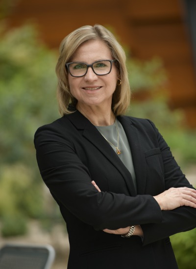
Dr. Russell Witte, a native Tucsonan, received a BS degree with honors in physics from the University of Arizona in Tucson (1993). Following travel abroad in Europe and Brazil, he began graduate studies in bioengineering at Arizona State University. His doctoral thesis (PhD, 2002) used chronic microelectrode arrays to describe sensory coding and learning-induced plasticity in the mammalian brain. He then moved to the University of Michigan in Ann Arbor and, as a post doc in the Biomedical Ultrasonics Laboratory, developed novel hybrid imaging techniques that integrate ultrasound, light, and/or microwaves for medical applications. In 2007, Dr. Witte returned to Tucson and is now Associate Professor of Medical Imaging, Optical Sciences and Biomedical Engineering at the University of Arizona. Dr. Witte’s Experimental Ultrasound and Neural Imaging Laboratory (EUNIL) devises cutting-edge imaging technology, integrating light, ultrasound and microwaves to diagnose and treat diseases ranging from chronic tendon disorders (tendinopathies) and irregular cardiac rhythms (arrhythmias) to breast cancer. By integrating different forms of energy, special effects are created that enable ultrasound imaging of optical absorption deep in tissue (photoacoustic imaging), mapping current source densities in the beating heart (acoustoelectric imaging), and elasticity imaging of human muscle and tendon for quantifying tissue mechanical properties. Dr. Witte's research further extends into nanotechnology and smart contrast agents, which have applications to functional brain imaging, cardiovascular disease, and cancer. Dr. Witte works closely with collaborators in the Colleges of Engineering, Optical Sciences and Medicine, as well as industry, to develop cutting-edge imaging technologies that potentially improve patient care. Dr. Witte is also a member of the Arizona Cancer Center, Sarver Heart Center and School of Mind, Brain, and Behavior, as well as the Neuroscience, Applied Mathematics, and Biomedical Engineering graduate interdisciplinary programs (GIDPs). Dr. Witte's vision is to develop a new generation of young investigators steeped in multiple disciplines branching from neuroscience, neural engineering, biochemistry, mathematics, biomedical imaging and, physics. He welcomes dreamers, brainstormers and problems solvers to join his team in search of the next great discovery in physics and medicine. Keywords: Biomedical Engineering/Medical Imaging


