Optical coherence tomography

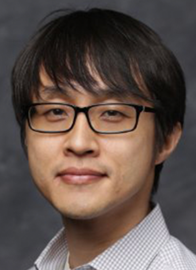
Dongkyun Kang

Assistant Professor, Optical Sciences
Assistant Professor, Biomedical Engineering
Assistant Professor, BIO5 Institute
Primary Department
Contact
(520) 621-6997

Work Summary

We are developing low-cost in vivo microscopy devices that can visualize cellular details of human tissues in vivo and help disease diagnosis and treatment in low-resource settings, high-speed tissue microscopy technologies that can examine entire organ under risk of having malignant diseases and detect small, early-stage lesions, and miniature microscopy devices that have the potential to examine anatomically-challenging human organs and facilitate integration of microscopic imaging with other imaging modalities.

Research Interest

My research is focused on developing novel optical microscopy technologies and improving patient care using these technologies. My research area includes (1) low-cost smartphone in vivo microscopy, (2) high-speed comprehensive in vivo endomicroscopy, and (3) ultraminiature endomicroscopy. (1) Low-cost smartphone in vivo microscopy: I am currently leading a NIH-sponsored research project for developing smartphone confocal microscope and diagnosing Kaposi's sarcoma in Uganda with the smartphone confocal microscope. I will further advance the smartphone microscopy technology and address other applications, including diagnosis of cervical and oral cancers in low-resource settings, large-population screening of skin cancers in the US, and aiding science and medical educations. (2) High-speed comprehensive in vivo endomicroscopy: I have previously developed a high-speed confocal microscopy system and endoscopic imaging catheters and acquired largest in vivo confocal images of human organ reported. At the UA, I plan to further advance the technology by i) increasing the imaging speed by orders of magnitude and ii) incorporating fluorescence imaging modality. (3) Ultraminiature endomicroscopy: In my previous research, I have developed miniature endoscopic catheters that can visualize internal organs in vivo through a needle-sized device. At the UA, I will develop microscopic imaging catheter with a extremely small diameter and utilize it for guiding cancer diagnosis and treatment.

Jennifer Kehlet Barton

Director, BIO5 Institute
Thomas R. Brown Distinguished Chair in Biomedical Engineering
Professor, Agricultural-Biosystems Engineering
Professor, Biomedical Engineering
Professor, Electrical and Computer Engineering
Professor, Medical Imaging
Professor, Optical Sciences
Professor, Cancer Biology - GIDP
Professor, BIO5 Institute
Member of the General Faculty
Member of the Graduate Faculty
Primary Department
Department Affiliations
Contact
(520) 626-0314

Work Summary

I develop new optical imaging devices that can detect cancer at the earliest stage. Optics has the resolution and sensitivity to find these small, curable lesions, and we design the endoscope that provide access to organs inside the body. .

Research Interest

Jennifer Barton, Ph.D. is known for her development of miniature endoscopes that combine multiple optical imaging techniques, particularly optical coherence tomography and fluorescence spectroscopy. She evaluates the suitability of these endoscopic techniques for detecting early cancer development in patients and pre-clinical models. She has a particular interest in the early detection of ovarian cancer, the most deadly gynecological malignancy. Additionally, her research into light-tissue interaction and dynamic optical properties of blood laid the groundwork for a novel therapeutic laser to treat disorders of the skin’s blood vessels. She has published over 100 peer-reviewed journal papers in these research areas. She is currently Professor of Biomedical Engineering, Electrical and Computer Engineering, Optical Sciences, Agriculture-Biosystems Engineering, and Medical Imaging at the University of Arizona. She has served as department head of Biomedical Engineering, Associate Vice President for Research, and is currently Director of the BIO5 Institute, a collaborative research institute dedicated to solving complex biology-based problems affecting humanity. She is a fellow of SPIE – the International Optics Society, and a fellow of the American Institute for Medical and Biological Engineering. Keywords: bioimaging, biomedical optics, biomedical engineering, bioengineering, cancer, endoscopes