
University of Arizona celebrates new AI-powered MRI and BIO5 Brain & Body Imaging Center
State-of-the-art technology and interdisciplinary expertise position the university at the forefront of imaging innovation in Arizona.
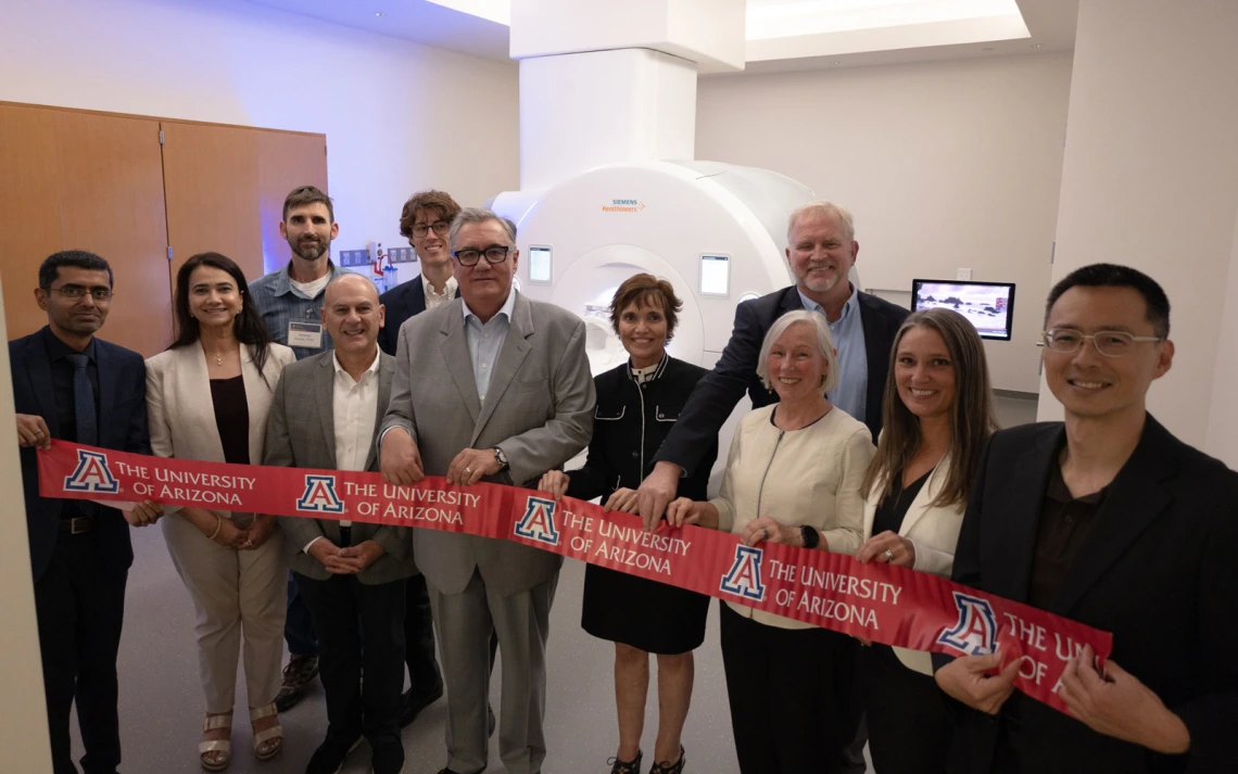
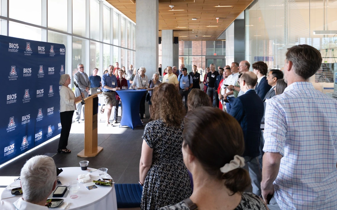
University of Arizona faculty and leadership celebrated new imaging technology with community and industry partners on September 3, 2025. Front row, from left: Vignesh Subbian, Hina Arif-Tiwari, CJ Karamargin (Congressman Juan Ciscomani), Tomás Díaz de la Rubia, Maria Altbach, Lee Ryan, Katie Grant (Siemens Healthineers), Nan-kuei Chen. Back row, from left: Andy Rouse, Berto Ibarra (Senator Ruben Gallego), Ted Trouard.
Deanna Rodriguez, BIO5 Institute
In early September, dozens of University of Arizona researchers and community leaders gathered to celebrate the installation of two of the most advanced MRI systems available today.
They represent a leap in non-invasive imaging. The AI-driven machines open new possibilities for treating neurological diseases like Alzheimer’s and Parkinson’s, as well as expanding the capabilities for imaging the heart, lungs, liver, even for patients with metallic implants.
“These MRI systems represent a strategic investment in technologies that harness artificial intelligence to improve human health,” said Tomás Díaz de la Rubia, senior vice president for research and partnerships. “By pairing these state-of-the-art tools with U of A's transdisciplinary expertise, we are accelerating innovation in discovery, diagnosis and treatment. This is how we deliver on the promise of AI-driven healthcare.”
Both MRIs are housed in the Translational Bioimaging Resource (TBIR), a university-wide core supported by the Office of Research and Partnerships (ORP). With expanded research facilities, TBIR now provides researchers across campus with broader access to advanced imaging tools and expertise.
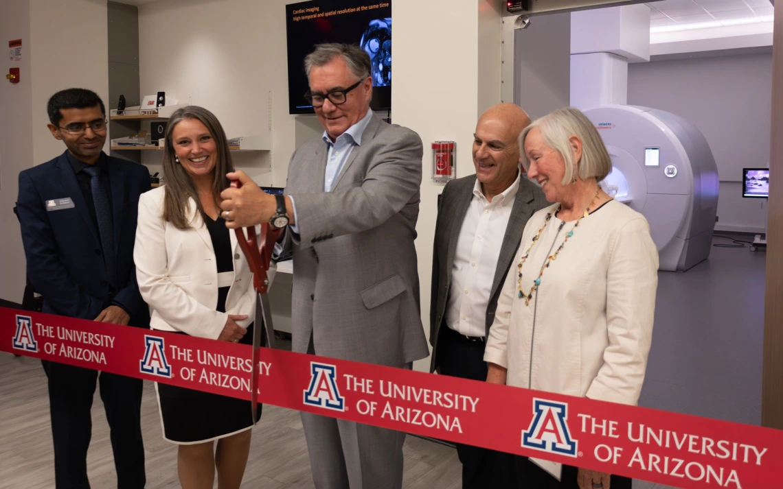
Tomás Díaz de la Rubia, senior vice president for research and partnerships, marked the arrival of advanced MRI technology with a ribbon-cutting in early September.
Deanna Rodriguez, BIO5 Institute
Building on that momentum, more than 30 researchers launched the new BIO5 Brain & Body Imaging Center (BBIC), uniting their expertise to maximize the impact of the new technology.
These milestones were made possible by federal and institutional investment, industry partnership, and years of cross-campus collaboration. That collective effort set the stage for this year’s celebration of two state-of-the-art MRI systems and the center.
Two new MRIs, one bold vision
A few years ago, the National Institutes of Health awarded University of Arizona researchers a $2 million grant to purchase a powerful new Siemens MRI for brain research. At the time, the university’s only research-dedicated MRI was booked solid, limiting opportunities for studies across campus.
In March 2025, the Siemens MAGNETOM Cima.X 3T—the company’s most advanced system for brain imaging—was installed.
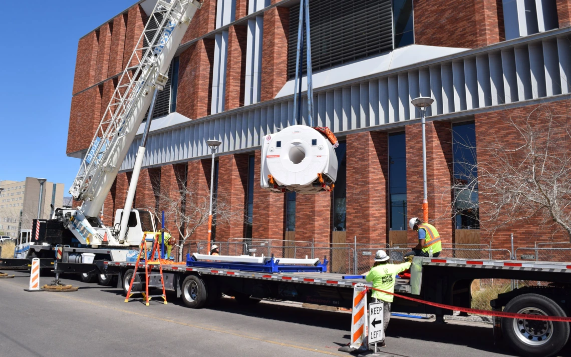
The Siemens Cima.X 3T was installed in March 25, 2025 at the Biosciences Research Laboratories on the University of Arizona campus.
Kristina Irwin, Brain & Body Imaging Center
Just months later, the Siemens MAGNETOM Free.Max 0.55T, designed to expand the capabilities of whole-body imaging, was added.
“What excites me most is knowing our researchers now have the very best possible MRI equipment so they can generate the best possible data for their studies” said Ted Trouard, professor emeritus of biomedical engineering and professor of medical imaging, and principal investigator on the NIH grant.
These milestones were made possible through federal investment from the National Institutes of Health, in partnership with Siemens Healthineers, and with support from the Office of Research and Partnerships, University of Arizona Health Sciences, the Colleges of Engineering, Medicine – Tucson, and Science, the University of Arizona Cancer Center, and the BIO5 Institute.
Together with existing TBIR technologies, the new MRIs bring unique yet complementary strengths to advance brain and body imaging.
AI at the core of imaging advances
Artificial intelligence drives both systems. It sharpens images, shortens scan times, and reveals more detailed patterns.
AI enhances MRI scans, revealing clearer details in less time.
Lily Howe, BIO5 Institute
These advances tie directly into the university’s broader AI + Health initiative to transform diagnosis, personalize treatment, and improve care across Arizona.
“AI is already making a huge difference in imaging, especially in how we reconstruct and analyze data,” said Lee Ryan, professor of psychology, radiology and imaging sciences, and neurology, and director of the BIO5 Brain & Body Imaging Center. “Both systems pair new hardware with AI-driven methods so we can see more, faster.”
Sharper brain imaging for Alzheimer’s and more
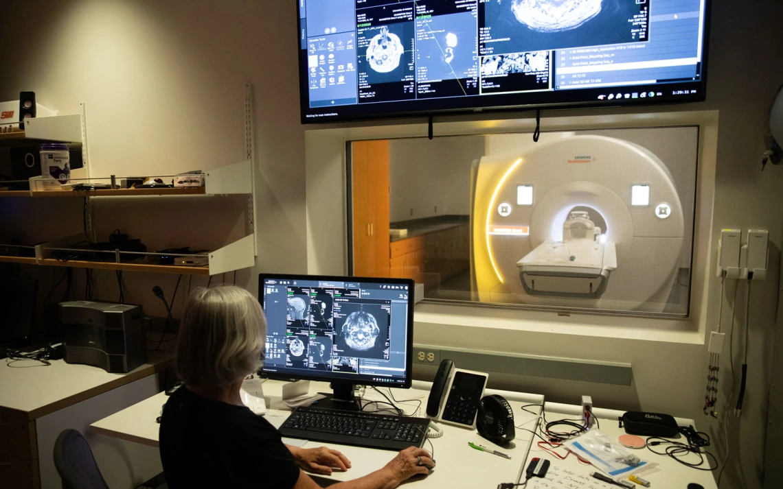
The Siemens Cima.X 3T is designed for brain imaging and produces ultra-high-resolution scans faster than ever before.
Building on the AI-driven advances already noted, the Cima.X 3T reduces scan time so patients spend less time in the machine while researchers collect more data.
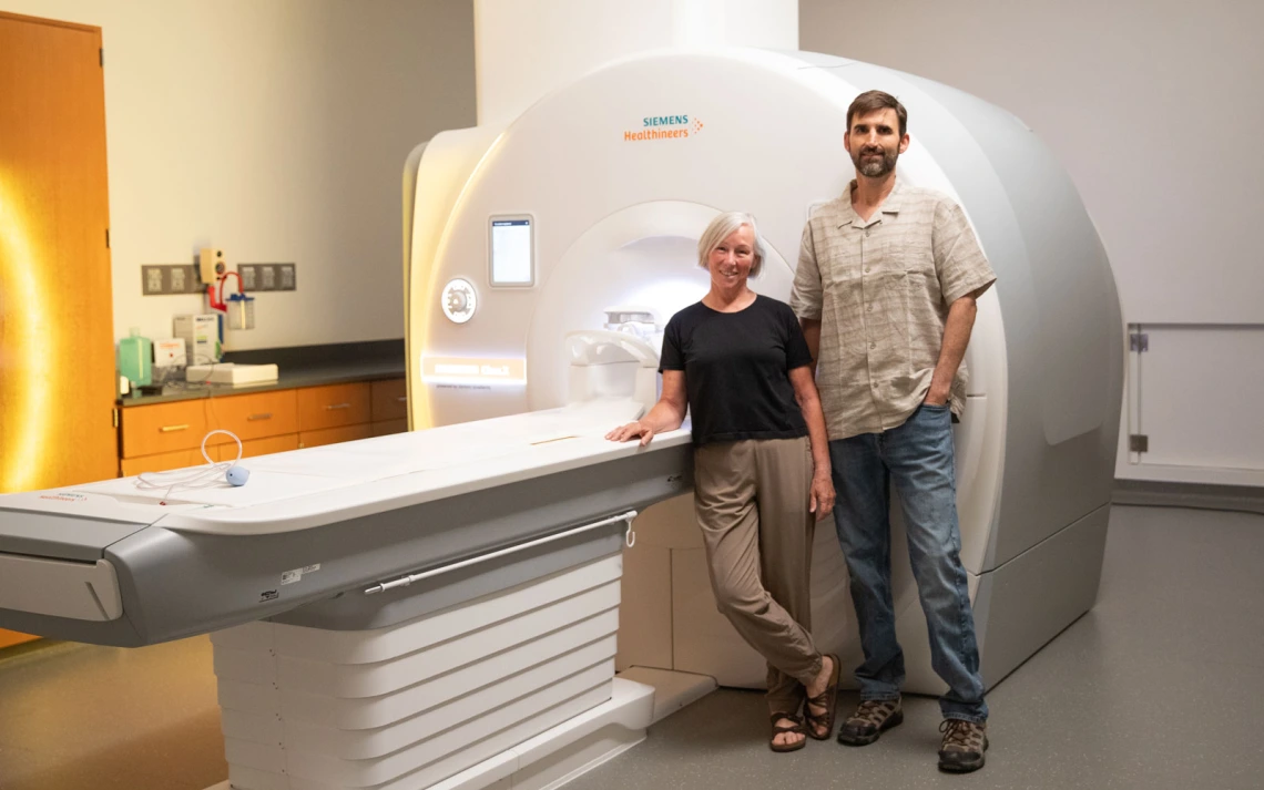
Lee Ryan, director of the BIO5 Brain & Body Imaging Center, and Andy Rouse, director of the Translational Bioimaging Resource, stand in front of the Siemens Cima.X 3T that delivers sharper, faster brain scans powered by AI.
Lily Howe, BIO5 Institute
One powerful example is a scan that tracks how water moves through brain tissue, helping researchers study connections and changes linked to Alzheimer’s, multiple sclerosis, and other neurological conditions.
Expanding access for body imaging
The Siemens Free.Max 0.55T has a lower magnetic field strength and a more open design, making it ideal for whole-body imaging.
Patients with metallic implants can be safely scanned, and the resulting images have minimal distortions due to metal. Its open design makes scans more comfortable and accessible, including for patients with larger bodies or during pregnancy.
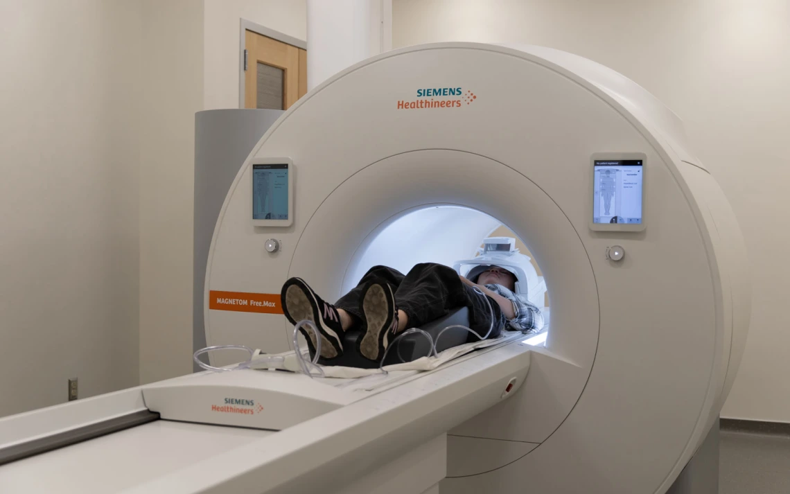
The open design of the Free.Max 0.55T eases the patient experience without sacrificing image quality.
Lily Howe, BIO5 Institute
AI reconstruction methods yield high quality images despite the lower field strength.
“Scanning at lower fields is advantageous for lung imaging. One project, recently funded by the Arizona Biomedical Research Centre, will use the Free.Max 0.55T and quantitative MRI methods developed by our team at the University of Arizona to look at the difference between Valley fever nodules and lung cancer,” said Maria Altbach, professor of radiology, imaging sciences, and biomedical engineering, and MRI expert on body imaging. “This could spare patients from invasive procedures like lung biopsies.”
The cost of the magnet and its installation is significantly lower compared to high field scanners, extending the capabilities of MRI to communities with limited resources.
These benefits highlight how technology improves patient care. But just as important is the scientific community that will harness these tools to push imaging research to new levels.
New hub for interdisciplinary imaging
Machines matter, but people make breakthroughs. While TBIR provides the equipment at the University of Arizona, BBIC brings together faculty, trainees, technicians, and industry experts and their knowledge to drive innovation.
“We’ve built a strong imaging community at the University of Arizona over the past several decades, and the Brain & Body Imaging Center gives us a centralized place to come together to share resources, apply for grants, and exchange interdisciplinary expertise,” said Ryan. “And I want to emphasize our name—our technology and people allow us to connect brain health to different parts of the body like never before.”
As part of the BIO5 Institute, the center strongly reflects the institute’s mission to connect disciplines and accelerate discovery.
“The BIO5 Brain & Body Imaging Center empowers ‘convergent science,’ where engineers, clinicians, neuroscientists, and data scientists innovate together on problems no single field could solve,” said Vignesh Subbian, associate professor of biomedical engineering and interim director of the BIO5 Institute.
Researchers across the University of Arizona campus are excited to explore the capabilities of the two new state-of-the-art MRI now available in the Biosciences Research Laboratories.
Deanna Rodriguez, BIO5 Institute
Equally important, the center will train the next generation of imaging scientists and clinicians in the latest technologies and AI-driven methods. It will not only accelerate current research but spark new questions and collaborations.
“We are always looking forward in imaging,” said Nan-kuei Chen, associate professor of biomedical engineering and expert in MRI physics. “These new technologies, along with the center, will allow us to expand into musculoskeletal imaging, sleep studies, and multi-modal brain research.”
The University of Arizona is expanding what’s possible in brain and body health research, by pairing advanced MRI technology with AI-driven methods and BIO5’s collaborative strength.